
- 14785 Jeffrey Road
- Suite 105
- Irvine, CA 92618
- Tel: 949.751.2089
- Fax: 949.502.6352
What is endodontics?
 Endodontics, a recognized specialty of the American Dental Association, focuses on the treatment of diseases that occur inside of the tooth, most commonly requiring conventional or surgical root canal therapy. Root canal treatment is a procedure to remove damaged tissue from the inside of the root canals of a tooth. When you look at your tooth in the mirror, what you see is the crown. The rest of the tooth, hidden beneath the gum line, is called the root. Inside the tooth, under the enamel and a hard layer called dentin, is a soft tissue called the pulp. The pulp contains connective tissue, blood vessels and nerves. As a result of deep decay, repeated dental procedures, periodontal disease, or tooth fractures, bacteria are introduced into the pulp and cause irreversible damage. When this happens, an endodontist removes the diseased pulp to save the tooth and prevent further infection and inflammation. After successful endodontic treatment, the tooth continues to perform normally.
Will I feel pain during or after the procedure?
The goal of endodontics is to relieve pain caused by pulp inflammation or infection. With modern anesthetic techniques, the majority of patients report that they are comfortable during the procedure. For the first few days after treatment, your tooth may be sensitive or sore, especially if there was pain or infection before the procedure. In the majority of cases, over-the-counter pain relievers are used for this discomfort, but your doctor may prescribe additional medications for you.
What happens after treatment?
 When your root canal therapy has been completed, a treatment report including digital images will be sent to your restorative dentist. You should contact your general dentist for a permanent restoration within a few weeks of completion at our office. Your restorative dentist will decide on what type of restoration is necessary to protect your tooth. We will contact you 3 to 12 months after treatment for a follow-up exam and digital image to monitor your healing.
Should I be worried about x-rays?
 We use digital radiography, an advanced computerized system that produces radiation levels 80 percent lower than conventional dental x-rays. These digital images can be optimized, archived, printed and sent to your general dentist via e-mail or CD.
What about infection?
Again, there's no need for concern. We adhere to the most rigorous standards of infection control advocated by OSHA, the Centers for Disease Control and the American Dental Association. We utilize autoclave sterilization and barrier techniques to eliminate any risk of infection.
What is an operating microscope?
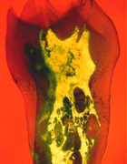
 In addition to digital radiography, we utilize special operating microscopes during your treatment. This technology allows the endodontist to magnify and illuminate deep into the root canals of a tooth, often visualizing the source of infection. The operating microscope can also be used to record images of your tooth and communicate additional information to your general dentist.
what is Cone Beam CT?
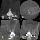
 In layman's terms, CBCT is a compact, faster and safer version of the regular CT. Through the use of a cone shaped X-Ray beam, the size of the scanner, radiation dosage and time needed for scanning are all dramatically reduced.
A typical CBCT scanner can fit easily into any dental ( or otherwise ) practice and is easily accessible by patients. The time needed for a full scan is typically under one minute and the radiation dosage is up to a hundred times less than that of a regular CT scanner. CBCT literally makes the tooth transparent in 3D so making the root canal anatomy, fractures, abscess, resorption and defects more detectable.
© 2024 Prestige Endodontics. All rights reserved.